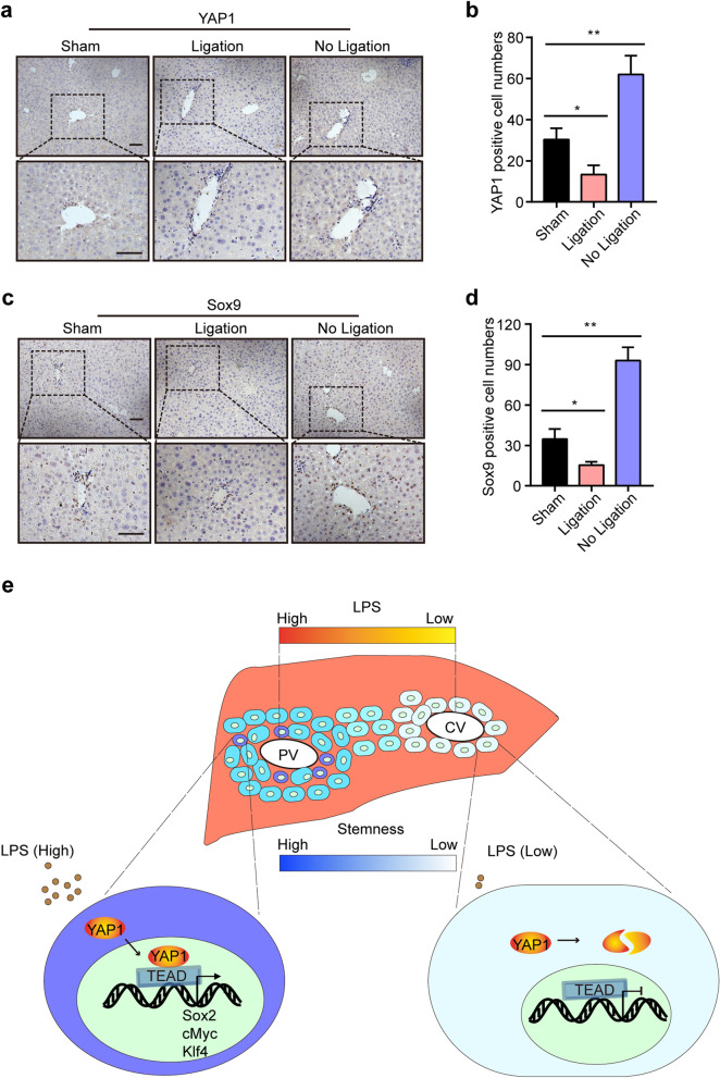
Fig. 7.
Correlation among LPS, YAP1, and Sox9+ stem cells in vivo. a, b Mice were administered a sham surgery or subjected to ligation of the left portal vein branch for 12 h and then sacrificed to harvest their liver lobes. IHC staining of YAP1 in the different liver lobes of WT mice in the sham and PVL groups (a). Quantification of the YAP1-positive cell numbers (b). Ligation: the left liver lobes with ligation of the portal vein from the PVL group; no ligation: the right liver lobes with no ligation of the portal vein from the PVL group. c, d IHC staining of Sox9 in the different liver lobes of mice in the sham and PVL groups (c) and the quantification of the Sox9-positive cell numbers (d). e Schematic representation of the LPS/YAP1 axis-mediated maintenance of hepatocyte stemness in the PV area. Scale bars, 50 μm. *P < 0.05, **P < 0.01

