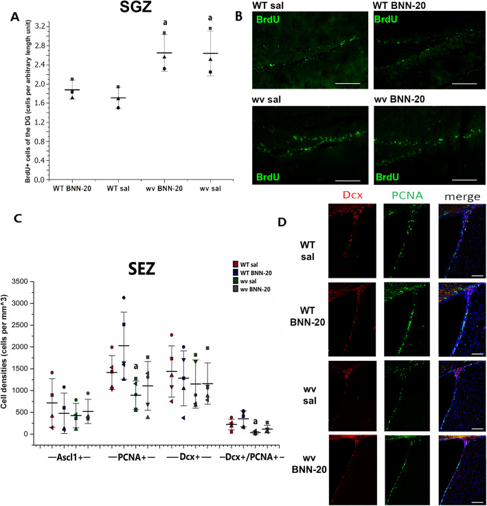
Fig. 4.
Lack of effect of BNN-20 administration on the main neurogenic zones of the postnatal brain. A Dot plot of numbers of BrdU+ cells per length and B representative immunofluorescence images of BrdU+ cells in the subgranular zone (SGZ) of the dentate gyrus (DG) of the hippocampus [n = 3 per group. Scale bar 200 μm. Error bars are SDs. a: p < 0.05 compared to the respective WT group, using two-way ANOVA (p = 0.002, F = 18.881) followed by LSD post hoc test]. C Histogram of the cell density of transient neural progenitors (Ascl1), proliferating cells (PCNA+), neuroblasts (Dcx+), and Dcx+/PCNA+ cells in the SEZ [n = 2 mice per group. a: p < 0.05 compared to WT sal. Analysis was performed by two-way ANOVA (PCNA+ comparison: p = 0.009, F = 8.902 for genotype, PCNA+/Dcx + comparison: p = 0.001, F = 18.030 for genotype) followed by LSD post hoc test. Error bars are SDs.]. D Representative immunofluorescence images of PCNA+ and Dcx+ cells within the SEZ of WT or wv mice post-administration of BNN-20 or saline (sal) during P14–P60 [scale bars = 200 μm]

