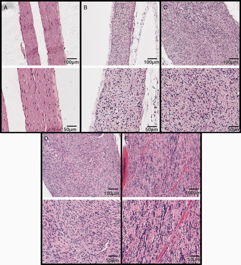
Figure 2.
Progressively increased atypia and hypercellularity within irradiated PNs after SI. H&E-stained sections of normal nerve and nerve sheath tumors arising after focal spinal irradiation. 10X magnification (top) and 20× (bottom) magnification images shown. (A) normal nerve from a wildtype mouse. (B) PN showing diffuse enlargement and replacement of the nerve fascicle by spindle cells and collagen. (C) CNF with more prominent hypercellularity. (D) ANNUBP. (E) Low-grade MPNST infiltrating skeletal muscle.

