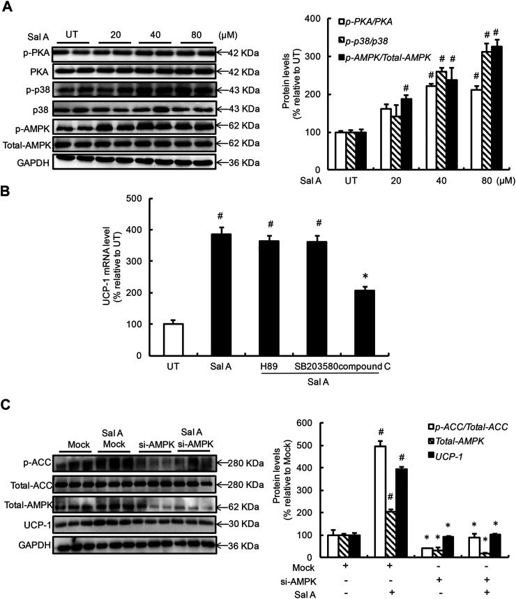
FIGURE 2.
AMPK activation contributes to Sal A-induced UCP-1 upregulation. (A) Differentiated C3H10T1/2 adipocytes were treated with Sal A (0, 20, 40, and 80 μM) for 4 h, intracellular p-PKA, PKA, p-p38, p38, p-AMPK, and AMPK contents were determined by Western blot. (B) Fully-differentiated C3H10T1/2 cells were pre-treated with PKA inhibitor (H89, 20 μM), p38 inhibitor (SB203580, 10 μM), and AMPK inhibitor (compound C, 1 μM), respectively, for 2 h before incubated with Sal A (80 μM) for 4 h. Intracellular UCP-1 mRNA level was measured by RT-PCR. (C) Fully-differentiated C3H10T1/2 adipocytes were transfected with siRNAs for AMPK. After silencing AMPK by siRNA, cells were exposed to 80 μM Sal A for 4 h. The protein levels of UCP1, AMPK, p-ACC and ACC were detected by Western blot. All values are denoted as means ± SD from three independent batches of cells. #p < 0.05 vs. the UT or Mock; *p < 0.05 vs. the Sal A treatment group. All groups contain two or three samples (n = 2 or n = 3).

