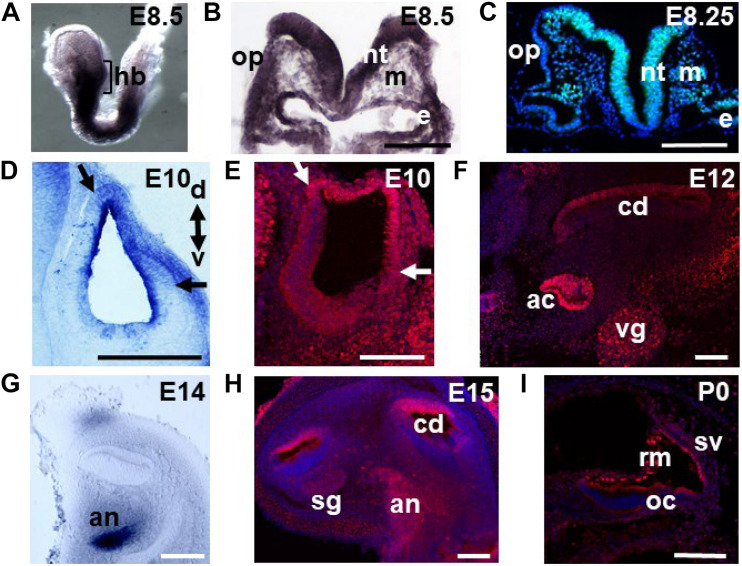
FIGURE 1.
Meis2 expression throughout inner ear development. Meis2 expression was detected via mRNA in situ hybridization (A,B,D,G) or immunohistochemistry (C,E,F,H,I) at the indicated stages. (A) At embryonic day (E) 8.5 high levels of Meis2 are detected at the level of the hindbrain (hb) and on corresponding sections at the level of the otic placode (op) (B). High levels of Meis2 protein are detected in the neural tube (nt), the endoderm (e), and weaker expression in the mesoderm (m) and the placode itself (C). (D) In the otic vesicle low Meis2 mRNA levels are observed in its dorsal-lateral portion (borders indicated by arrows). The orientations of the sections through the otic vesicles along the dorsal (d)–ventral (v) axis are indicated. (E) Immunohistochemical detection of Meis2 protein in the dorsal-lateral quadrant of the otic vesicle. (F) Presence of Meis2 protein in the roof of the cochlear duct (cd), the vestibular ganglion (vg) and the ampullary cristae (ac). (G) High levels of Meis2 mRNA are detected in the auditory nerve (an). (H) Next to the auditory nerve, Meis2 protein is also observed in the roof of the cochlear duct and to a lesser extent in the spiral ganglion (sg). (I) At P0 Meis2 immunoreactivity is detected in Reissner’s membrane (rm) and the stria vascularis (sv). oc, organ of Corti. Scale bars in (B,C,E–H): 100 μm; in (D): 200 μm and in (I): 75 μm.

