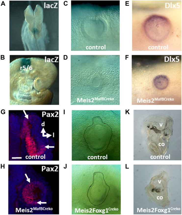
FIGURE 3.
Effects of tissue-specific inactivation of Meis2 on inner ear formation. (A,B) lacZ ROSA26 reporter staining caused by the MafBCre driver in the posterior hindbrain of E8.25 embryos and in rhombomeres (r) flanking the otic vesicle at E9 (B). (C–H) Meis2flox/flox; MafBCre/+ mutants show reduced sized vesicles (C,D) which maintain expression of Dlx5 (at E9) and Pax2 (at E9,5), as revealed by whole mount RNA in situ hybridization (E,F) and immunohistochemistry (G,H). The orientations of the sections through the otic vesicles along the dorsal (d)–lateral (l) axis are indicated. (I–L) Bright field view of otic vesicles at E11.5 and cleared inner ears at P1 from Meis2flox/flox; Foxg1Cre/+ mutants. Note the flask-shaped morphology of the otic vesicle and the lack of a discernible cochlea (co) in the mutants. v, vestibule.

