Fig. 4.
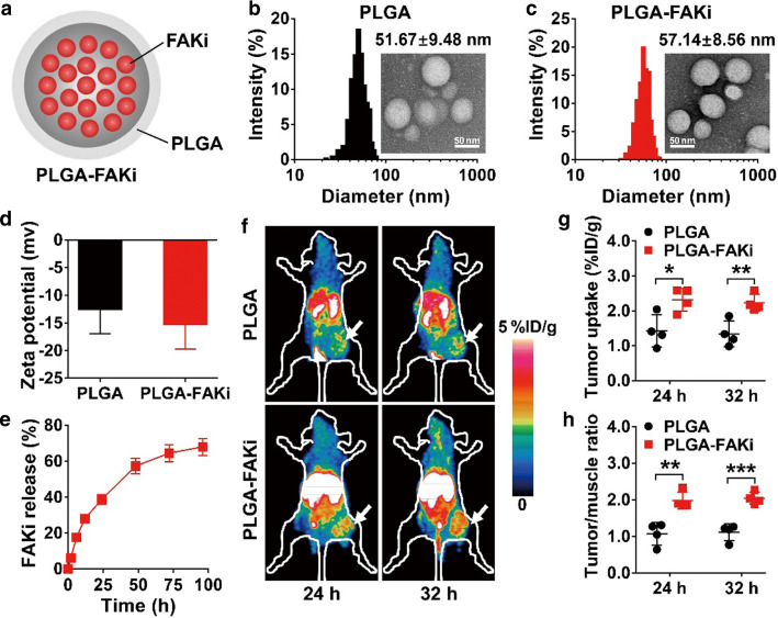
PET imaging of 64Cu-labeled OVA-specific cytotoxic T lymphocytes (OVA-CTLs) in B16-OVA tumor-bearing C57BL/6 mice treated with PLGA nanoparticles encapsulated with PF-562271 (PLGA-FAKi) or the control PLGA nanoparticles. a Schematic illustration of PLGA-FAKi. b, c Transmission electron microscope images and hydrodynamic diameters of PLGA (b) and PLGA-FAKi (c). d Surface zeta potentials of PLGA and PLGA-FAKi (n = 3). e Time-dependent profile of the release of PF-562271 (FAKi) from PLGA-FAKi nanoparticles dispersed in PBS at 37 °C (n = 3). f–h Small-animal PET images (f), quantified tumor uptake (g), and quantified tumor-to-muscle ratios (h) of 64Cu-OVA-CTLs at 24 and 32 h postinjection in B16-OVA tumor-bearing C57BL/6 mice after treatment with the control PLGA or PLGA-FAKi (n = 4/group). Tumors are indicated by white arrows. Data are presented as mean ± SD. *, P < 0.05; **, P < 0.01; ***, P < 0.001

