Fig. 1.
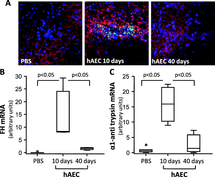
hAEC engrafted in the liver and expressed FH and α1 anti-trypsin mRNA. A Representative images of HNA+ (green) and DAPI+ (blu) cells in livers from Cfh−/− mice given either PBS or hAEC injection and euthanized 10 or 40 days later. No HNA-positive hAEC were detected in the livers of mice given PBS. The liver sections were counterstained for hepatocyte antigen expression (red staining). Original magnification × 400. mRNA expression of human FH (B) and anti-trypsin (C) in the livers of Cfh−/− mice given either PBS or hAEC injection and euthanized 10 or 40 days after. Horizontal lines indicated statistically significant differences (P < 0.05) between groups

