Fig. 1.
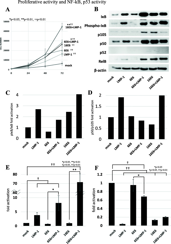
Cell proliferation, NF-κB activity and p53 induction. A Cell proliferation was compared among MEFs expressing viral proteins. The cells co-expressing low- or high-risk HPV E6 + EBV LMP-1 (6E6 + LMP-1 and 16E6 + LMP-1) grew faster than those expressing single viral protein and mock cells. Asterisk symbol indicates significant increase in cell number compared with cells expressing HPV E6 alone (*p < 0.05, **p < 0.01). Dagger symbol indicates a significant increase in cell number compared with mock cells (††p < 0.01). B Low-risk HPV6 E6 + EBV LMP-1 (6E6 + LMP-1) showed phosphorylation of IκB, processing of p105 to p50, and high RelB expression which were comparable to those seen in high-risk HPV16 E6 and EBV LMP-1 (16E6 + LMP-1). C and D) Relative pIκB/IκB (C) and p50/p105 (D) ratios of each clone were determined by densitometry. The ratios in mock cells were set to 1.0. Although high-risk HPV16 E6 + EBV LMP-1 (16E6 + LMP-1) showed increased ratios of both pIκB/IκB and p50/p105, low-risk HPV6 E6 + EBV LMP-1 (6E6 + LMP-1) had a high pIκB/IκB ratio but a p105/p50 ratio comparable to mock cells. E A luciferase assay for NF-κB activity indicated that low-risk HPV6 E6 + EBV LMP-1 (6E6 + LMP-1) had higher activity than cells expressing HPV6 E6 alone (6E6). However, high-risk HPV16 E6 + EBV LMP-1 (16E6 + LMP-1) showed more than eight-fold increase in activity over low-risk HPV6 E6 + EBV LMP-1 (6E6 + LMP-1). Asterisk and dagger symbols indicate a significant increase of NF-κB activation compared with cells expressing HPV E6 alone (*p < 0.05, **p < 0.01) and mock cells (†p < 0.05, ††p < 0.01), respectively. F Induction levels of p53 of each clone were compared through a luciferase assay. p53 expression decreased more in low-risk HPV6 E6 + EBV LMP-1 (6E6 + LMP-1) than those expressing HPV6 E6 alone (6E6). However, p53 suppression in low-risk HPV6 E6 + EBV LMP-1 (6E6 + LMP-1) was lower than that in high-risk HPV16 E6 + EBV LMP-1 (16E6 + LMP-1). Asterisk and dagger symbols indicate a significant decrease of p53 activation compared with cells expressing HPV E6/E7 alone (*p < 0.05) and mock cells (†p < 0.05, ††p < 0.01), respectively

