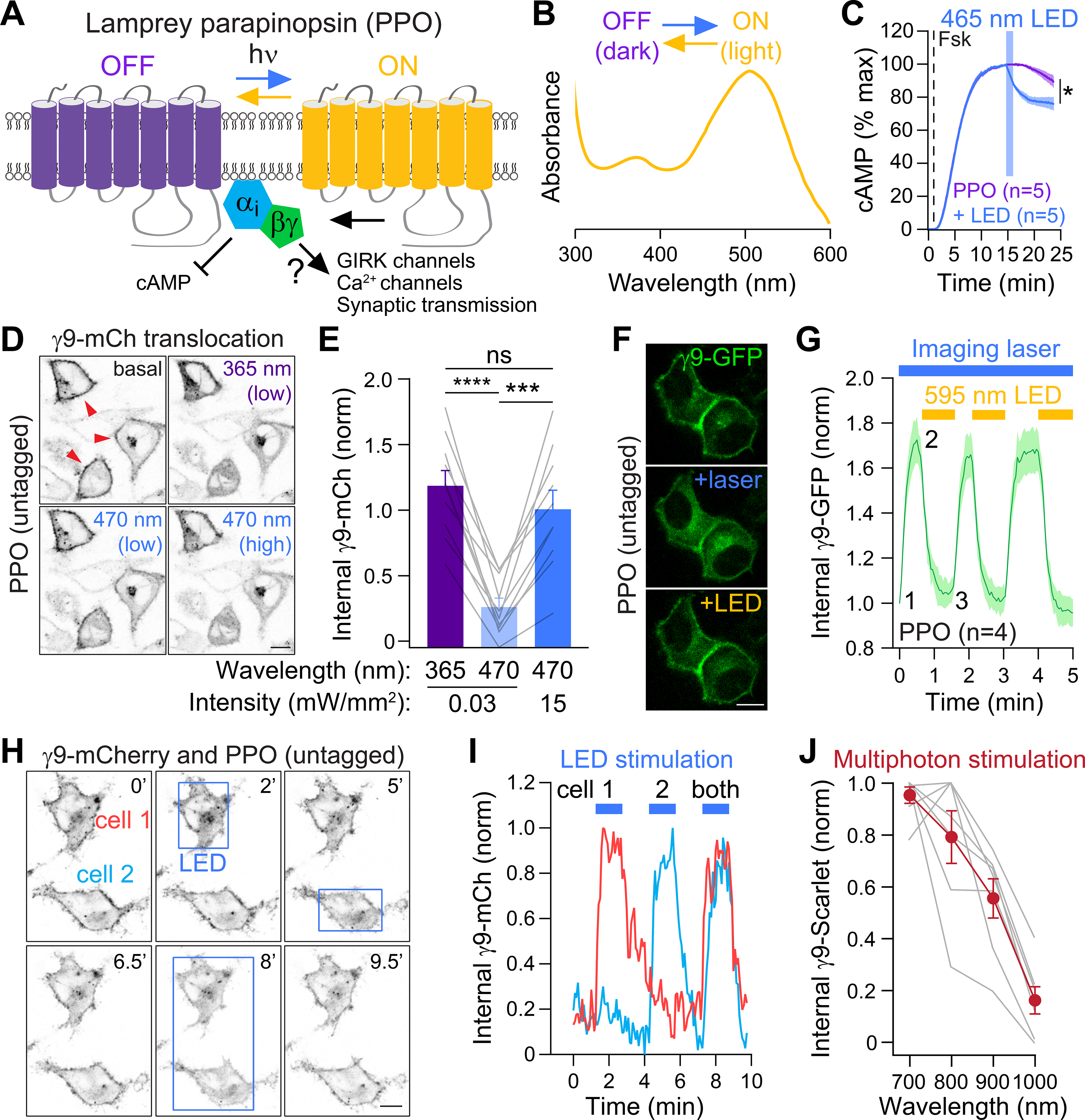
Figure 1. Spectral characterization of a photoswitchable GPCR-based opsin.

(A) Cartoon of parapinopsin (PPO), a photoswitchable GPCR that is activated by UV light and turned off by amber light.
(B) Absorption spectra of purified PPO protein in the dark off state (purple) and after UV light (amber). Graph is modified from (Koyanagi et al., 2004) (copyright 2004 National Academy of Sciences, USA)
(C) Optical stimulation with blue LED light (465 nm, 15.6 mW/cm2) inhibits forskolin-induced cAMP luminescence in PPO-expressing HEK cells. n=5
(D) Live-cell images of Gβγ translocation assays. In the dark γ9-mCherry is localized to the plasma membrane (PM) (red arrows) but translocates after stimulation of PPO with UV (365 nm) or blue (470 nm) light. Scale=10 μm
(E) Quantification of γ9-mCherry translocation following stimulation with UV vs. blue light. n=10, Paired t-test, ns=not significant, ***p<0.001, ****p<0.0001
(F) Translocation assays of PPO photoswitching. GFP-γ9 was imaged every 3s with a 488 nm laser (~45 μW), which also activated PPO and caused GFP-γ9 translocation from the PM (+laser). This was reversed by widefield illumination with an amber LED (590 nm, ~150 μW). Scale=10 μm.
(G) Quantification of γ9-GFP translocation. n=4 cells
(H) Individual photoactivation of cells expressing γ9-mCherry and untagged PPO. Boxes depict regions of blue LED photoactivation. Scale=10 μm
(I) Traces of γ9-mCherry translocation from panel H.
(J) Multiphoton activation of PPO using γ9-translocation assays. γ9-mScarlet was imaged with a Ti:Sapphire laser at 1080 nm, and multiphoton stimulation was delivered by a second laser at 700–1000 nm. n=7
