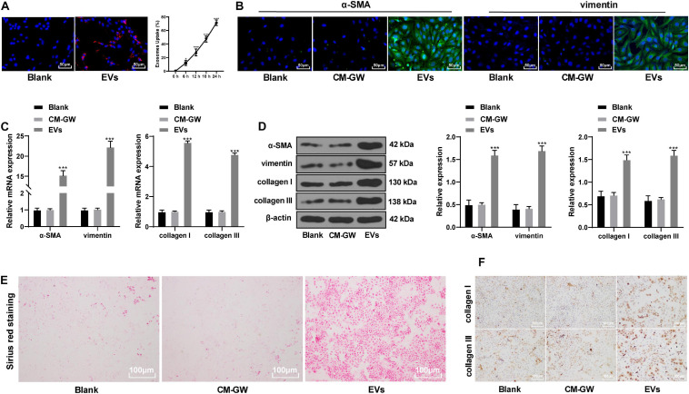
FIGURE 2.
EVs derived from hepatoma cells promoted the differentiation of BMSCs into fibroblasts. (A) Dil was used to label the EVs and to find the internalization of the EVs by BMSCs. Compared with 0 h, *p < 0.05, ***p < 0.001. (B) The expression of α-SMA and vimentin in BMSCs was detected by immunofluorescence. The stronger the green fluorescence, the higher the positive expression. (C,D) The levels of α-SMA, vimentin, collagen I, and collagen III in EVs-treated BMSCs were detected by RT-qPCR and Western blot analysis; compared with the blank group, ***p < 0.001. (E) Picric acid-Sirius red staining measured the collagen deposition in BMSCs. (F) levels of type I collagen and type III collagen in EVs-treated BMSCs were measured by immunocytochemistry. Data were analyzed by one-way ANOVA, followed by Tukey’s multiple comparison test. Replicates = 3.

