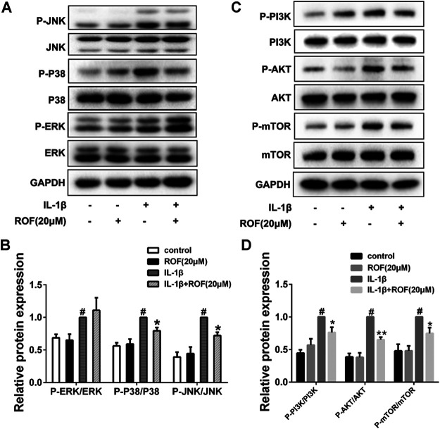
FIGURE 5.
ROF blocks the activation of P38/JNK and PI3K/AKT/mTOR pathways induced by IL-1β in chondrocytes. Cells were exposed to L-1β (10 ng/ml) with or without 20 μM ROF for 30 min as above. (A) Western blots and (B) quantitative analysis of MAPK pathways in each group. (C) Western blots and (D) quantitative analysis of PI3K/AKT/mTOR pathways in each group. JNK, P38, ERK, PI3K, AKT, and mTOR were used as loading control (n = 3). #p < 0.05 vs. control group; *p < 0.05 and **p < 0.01 vs. IL-1β group.

