Figure 4.
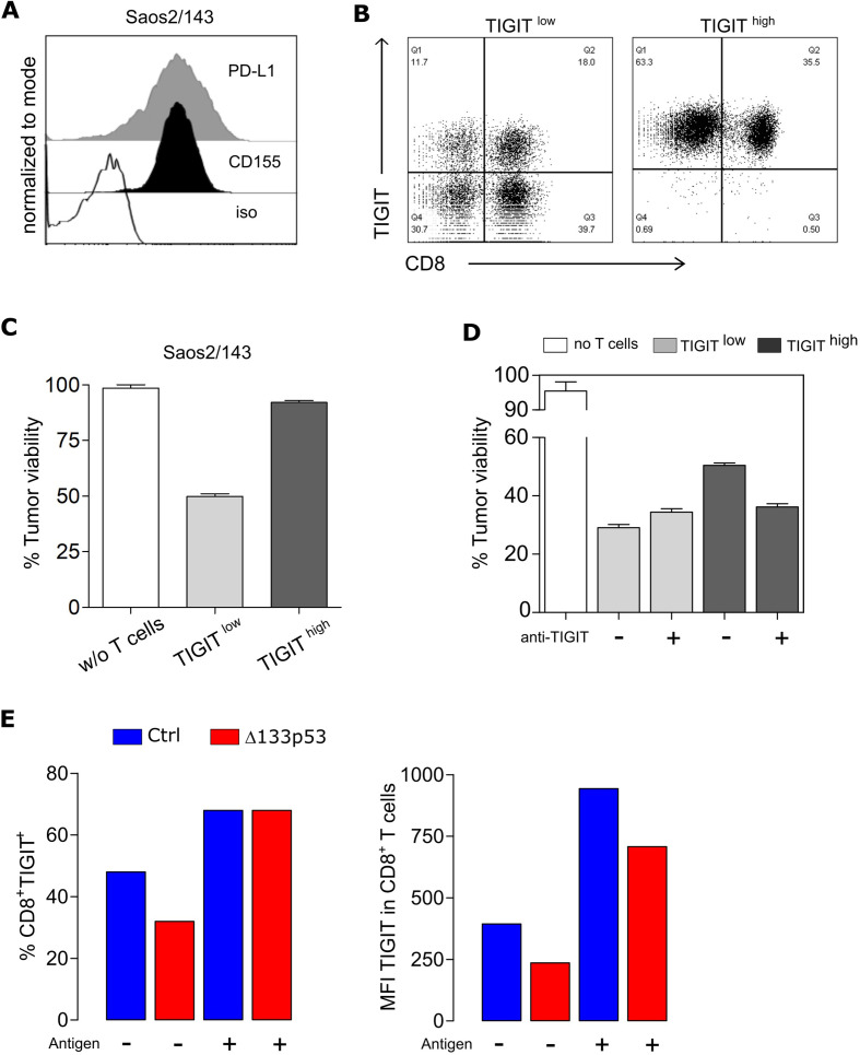
TIGIT induction is antigen-dependent and affects TCR-mediated T cell cytolytic activity. (A) Expression levels of PD-L1 and CD155 in the target tumor cell line Saos2/143 determined by flow cytometry. (B) Flow Cytometry Data showing TIGIT expression of CD8+ T cells after MACS-separation into TIGITlow and TIGIThigh population. (C) Quantified killing capacity of TIGITlow and TIGIThigh T cells was determined in a tumor colony-forming assay, and is expressed as the percentage of remaining viable tumor cells after coculture with effector T cells. (D) Quantified killing capacity of TIGITlow and TIGIThigh T cells on TIGIT blockade as determined in a tumor colony-forming assay. (E) Upregulation of TIGIT on CD8+ T cells before and after antigen recognition via coculture with Saos2/143. Percentage (left) and MFI (right) was assessed by flow cytometry. Results from one representative experiment out of three biological replicates are shown. MFI, mean fluorescence intensity; PD-L1, programmed cell death ligand 1.

