
Fig. 2.
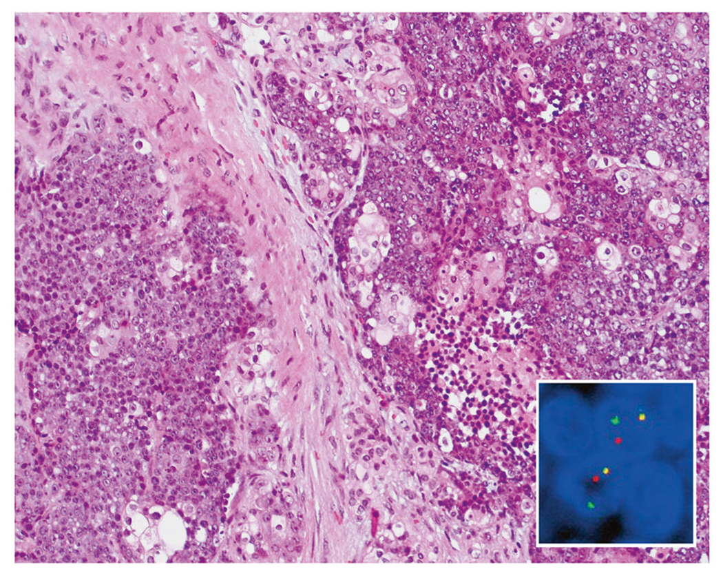
NUTM1-rearranged neoplasia. Typical examples of NUT carcinoma consist of primitive cells with variable foci of “abrupt” keratinization (hematoxylin and eosin, ×100). Break-apart NUTM1 FISH probes show split green and orange signals in case 26, indicating a NUTM1 gene rearrangement (inset)