
Fig. 3.
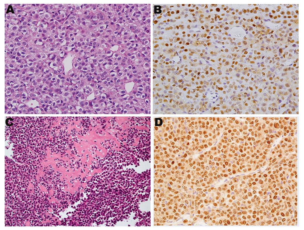
NUTM1-rearranged tumors with variant morphology. a, b Case 26 showed undifferentiated round cells along with NUT expression (NUT IHC in b). This case harbored NUTM1 gene break by break-apart FISH, leading to a diagnosis of NUT-rearranged tumor. c Case 22 showed sheets of epithelioid to plasmacytoid cells with production of dense extracellular collagen type material and harbored an MXD4-NUTM1 fusion. d Case 22 showed nuclear expression of NUT. (a, c, hematoxylin and eosin, ×400 and ×200, respectively; b, d, ×400)