
Figure 3.
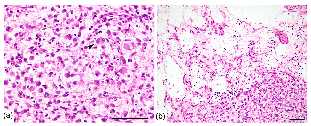
Histological findings. (a) The tumor consisted of scattered middle epithelioid tumor cells with abundant eosinophilic cytoplasm and moderately enlarged vesicular nuclei within a loose myxoid background. A binucleated tumor cell (arrow head) was present. (b) Liquefactive necrosis with lymphoid infiltration was observed. Bar, 100 μm.