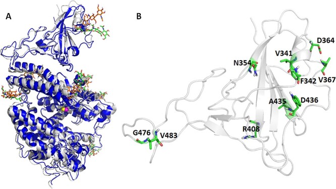
Figure 1 .

Overlay of crystal and MD representative structure for the wild type RBD/hACE2 (Panel A) and the 10 RBD mutation sites (Panel B). Glycosylated residues are shown in greenish sticks for the X-ray and brownish sticks for representative MD structures. The residues undergo mutations are shown as greenish sticks.
