FIGURE 4.
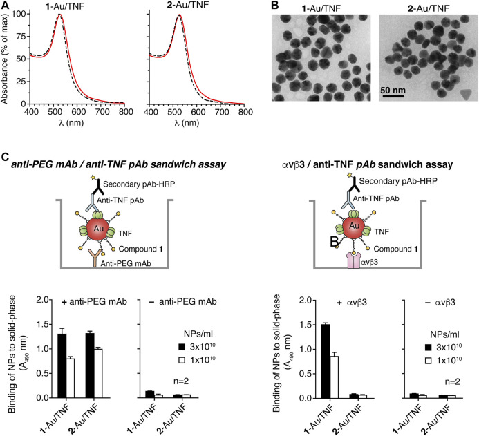
Characterization of 1-Au/TNF and 2-Au/TNF nanoparticles. (A) UV-Vis absorption spectra of nanogolds. The dotted line corresponds to uncoated 25 nm gold nanoparticles. (B) Transmission electron microscopy (TEM) of 1-Au/TNF and 2-Au/TNF. Representative microphotographs of 1-Au/TNF and 2-Au/TNF are shown. Morphometric analysis of nanoparticles shows that 1-Au/TNF and 2-Au/TNF consist of gold nanospheres with maximal diameters of 25.6 ± 2.3 nm and 26.7 ± 2.2 nm (mean ± SD, n = 100 NPs), respectively, and with a roundness value of 0.87 ± 0.07 and 0.090 ± 0.05, respectively (a roundness value of 1 corresponds to a perfect circle). (C) Binding of nanoparticles to microtiter plates coated with (+) or without (–) an anti-PEG mAb or αvβ3 integrin, as detected with an anti-TNF polyclonal antibody and HRP-labeled goat anti-rabbit antiserum. The results of a representative experiment are shown. Bars, mean ± SE of technical duplicate.

