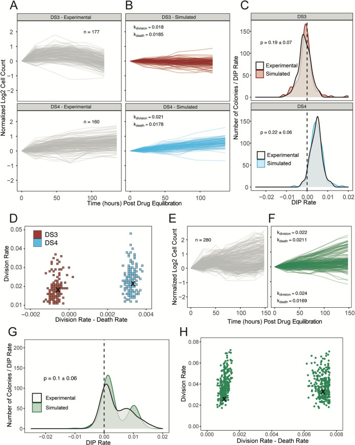
Fig 5. Stochastic simulations of a simple birth-death model reproduce DIP rate distributions from PC9-VU sublines.
(A) Experimental cFP time courses for 2 representative sublines (DS3 and DS4) in response to 3 μM erlotinib (same data used to generate the corresponding DIP rate distributions in Fig 2D). Each trace corresponds to a single colony, normalized to 72 h postdrug treatment. Only colonies with initial cell counts greater than 50 at the time of treatment are shown. (B) In silico cFP time courses from a one-state model with division and death rate constants that closely reproduce the experimental time courses in A. Trajectories are normalized to the time at which the simulated drug treatment was initiated, simulated cell counts are plotted only at experimental time points, and only colonies with initial cell counts greater than 50 at the time of simulated drug treatment are shown. (C) Comparison of experimental and simulated DIP rate distributions from time courses in A and B. Distributions are compared statistically using the AD test [66]. Dashed black line signifies zero DIP rate, for visual orientation. (D) Parameter scan of division and death rate constants for the 2 sublines in A–C. For each pair of rate constants, the same number of model simulations were run as the associated experimental cFP time courses in A. DIP rates were calculated and compiled into a distribution and then statistically compared against the corresponding experimental DIP rate distribution using the AD test. All parameter pairs with p < 0.05 (see Materials and methods) are colored white, indicating lack of statistical correspondence to experiment. “×” denotes a division and death rate constant pair used in B. (E) Same as A but for subline DS8. (F) Same as B but for DS8 using a two-state model. (G) Same as C but for DS8. (H) Same as D but for DS8 using a four-dimensional (2 division-death rate constant pairs) parameter scan and projected into 2 dimensions. “×” denotes parameter values used to generate the simulated DIP rate distribution in F. The data underlying this figure can be found in github.com/QuLab-VU/GES_2021. AD, Anderson–Darling; cFP, clonal fractional proliferation; DIP, drug-induced proliferation.

