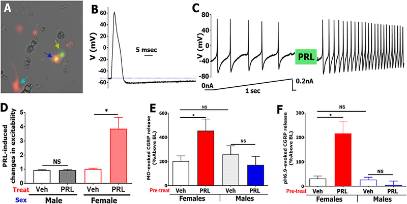
FIGURE 5:
PRL selectively sensitizes Prlr-positive TG neurons innervating the dura of female mice. Prlr-cre+/WGA-488+ back-traced neurons from dura TG neuron (blue arrow) were selected for recording. Yellow arrow shows Prlr-cre-/WGA-488+ TG neuron. Sapphire arrow shows Prlr-cre+ non-neuronal cell. (A) Action potential (AP) from small-sized (25 pF) selected Prlr-cre +/WGA-488+ TG neuron. Stimulus waveform is 1,000 pA, 0.5 ms. (B) Train of APs in a selected Prlr-cre+/WGA-488+ TG neuron (same neurons as panels A and B) was stimulated with current ramp protocol shown below trace. The neuron was treated with exogenous PRL (200 ng/ml) for 5 min, and the same current ramp protocol was applied. Ratio of post-PRL AP frequency to before-PRL AP frequency reflects changes in excitability. (C) PRL-induced changes in excitability of Prlr-cre+/WGA-488+ TG neuron from females and males. Control is vehicle treatment between two current ramps, n = 7–14 (D) PRL (1 μg/ml)-induced sensitization of MO (0.01%)-evoked CGRP release from female and male dura biopsies, n = 4–6. (E) PRL (1 μg/ml)-induced sensitization of pH 6.9-evoked CGRP release from female and male dura biopsies, n = 4 (F) Statistics are 2-way ANOVA with variables as sex and treatment (NS, non-significant; *p < 0.05).

