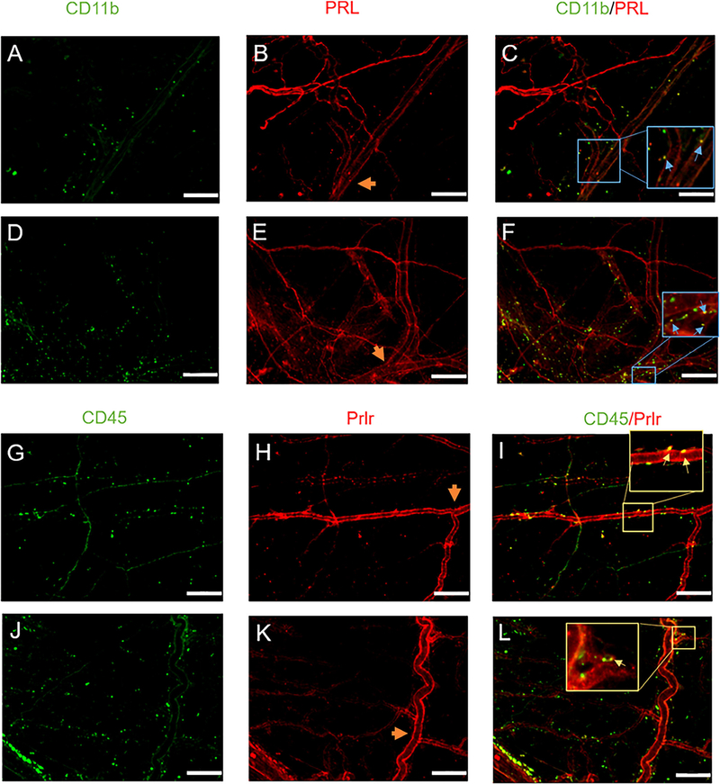
FIGURE 7:
Expression of prolactin and prolactin receptor in non-neuronal cells in the dura. Dura mater was removed from 3–5-month-old female (A-C) and male (D-F) mice. These dura were processed and stained for PRL expression and co-stained with CD11b. Images were taken at 20x. Scale bars represent 100 μm. Blood vessels are included as a potential source of prolactin release. Orange arrow heads indicate the middle meningeal artery (MMA) which served as a biological marker to ensure images were taken from the same region of animals. CD11b expression (A, D), PRL expression (B, E) and the overlay is shown in (C, F). Inserts in (C, F) highlight overlap of PRL and CD11b. Expression of Prlr in intact females. Prolactin receptor expression (H) was assessed in 3–5-month-*old female mice, dura was co-stained for CD45 (G) and overlap shown in (I). Dura of littermate Prlr conditional knockout animals, that have Prlr deleted from Nav1.8 sensory neurons, were also stained to assess the presence of Prlr (J-L). Inserts in (I, L) highlight overlap of Prlr and CD45.

