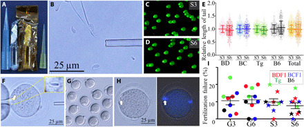
Fig. 3. Examination of sperm damage.

(A) Ampule of space-preserved FD sperm. (B) After rehydration, some of the sperm heads separated from the tails. (C and D) Comet DNA breakage assays of 2 years and 9 months (S3) and 5 years and 10 months (S6) space-preserved FD sperm. Two independent technicians measured the straight lengths from sperm heads (bright spot) to the edges of the faint light. (E) The lengths of DNA in the comet tails were standardized against the average lengths of the S3 FD sperm results for each mouse strain. (F) FD sperm were injected into fresh BDF1 oocytes. Arrows indicate injected sperm heads. (G) Six hours later, most of the oocytes were fertilized. (H) Some of the oocytes failed to fertilize after FD sperm injection, and those oocytes had pseudo-MII spindles derived from sperm nuclei [left, bright image; right, DNA under ultraviolet (UV) light]. (I) The rate of fertilization failure between preservation periods and between mouse strains. Key: mouse strains: BD, BDF1; BC, BCF1; Tg, B6129F1-GFP; B6, C57BL/6. Photo credits: (A, B, and F to H) Sayaka Wakayama and (C and D) Daiyu Ito, University of Yamanashi.
