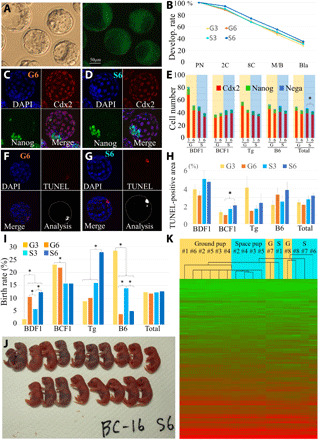
Fig. 5. Examination of embryo quality and full-term development.

(A) Blastocysts derived from embryos fertilized with space-preserved spermatozoa of 129B6F1-Tg strain (left) and GFP expression under UV light (right). (B) Developmental rate of embryos up to blastocyst stage. PN, pronuclear stage; 2C, two-cell stage; 8C, eight-cell stage; M/B, morulae/blastocyst stage; Bla, blastocyst stage. Immunostaining of blastocysts fertilized with G6 ground control (C) and space sperm (D). The nuclei of embryos were detected by nuclear staining using DAPI (top left, blue). CDX2-positive cells [trophectoderm (TE)] are red (top right), Nanog-positive cells [inner cell mass (ICM)] are green (bottom left), and merged images (bottom right). The cell number of TE, ICM, and both negative Ne are shown in (E). The incidence of apoptosis in blastocysts derived from G6 ground control (F) or space sperm (G) were examined using the TUNEL assay. The foci of TUNEL signals show apoptosis cells (top right, red). Merged images (bottom left) and converted digital images using ImageJ software (bottom right). (H) The red area of each blastocyst was plotted. (I) Full-term development of embryos fertilized with G3 and G6 ground control and S3 and S6 space-preserved spermatozoa. Asterisks show significant differences (P < 0.05). (J) 19 pups were obtained from S6 space-preserved FD sperm from one experiment. Key for mouse strains: B6, C57BL/6N; BD, B6D2F1; BC, B6C3F1; Tg, 129B6F1 expressing GFP. (K) The results of the RNA sequencing (RNA-seq) analysis are shown as heatmaps. Photo credits: (A) Teruhiko Wakayama, (C and D) Kosuke Kazama, (F and G) Naoki Hirose, and (J) Sayaka Wakayama, University of Yamanashi.
