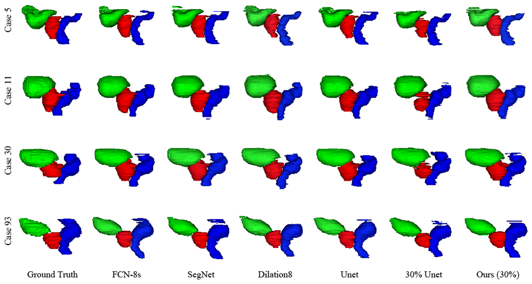
Fig. 5.

3D visualization of the segmentation results of the prostate, bladder, and rectum obtained by the compared FCN approaches trained using the completely annotated data and the incompletely annotated data with 30% ground-truth labels, respectively. Four rows indicate the results of four patients. Red denotes the prostate, green denotes the bladder, and blue denotes the rectum.
