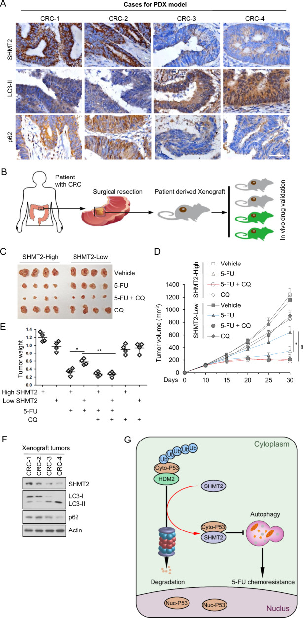
Fig. 6. CQ sensitizes PDXs with low SHMT2 expression to 5-FU treatment.
A Images of immunohistochemical staining for SHMT2, LC3, and p62 in CRC tissues from four selected patients (two with low SHMT2 expression and two with high SHMT2 expression) using the indicated antibodies. Scale bar, 50 μm. B Schematic of PDX model establishment. C–E Xenograft experiments with 5-FU or CQ treatment are described in the Methods section. C Xenograft tumors were harvested and photographed. D, E Quantification of the average volumes (D) and weights (E) of the xenograft tumors are shown. Four tumors from individual mice were included in each group; *P < 0.05, **P < 0.01. F Representative western blot of xenograft tumors. G Schematic diagram showing the basic hypothesis/conclusion/model.

