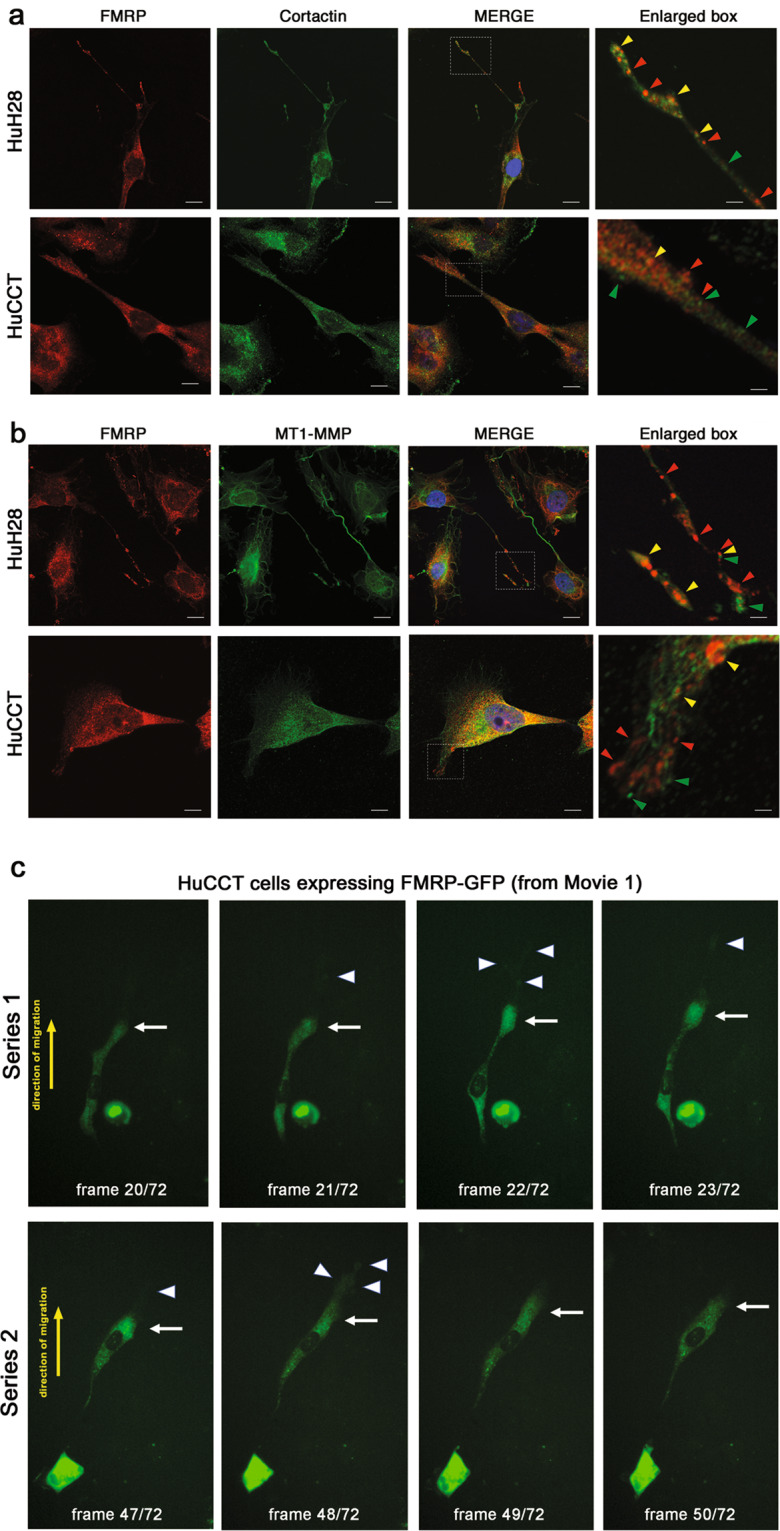
Fig. 3. FMRP localizes in cytoplasmic granules inside Cortactin and MT1-MMP –enriched cell protrusions.

a HuH28 or HuCCT cells were grown on fibronectin-coated chamber slides for 48 h and then IF was performed. Cells were stained with FMRP (red), Cortactin (green) and nuclei were stained with DAPI (blue). In the last right panels, enlarged images of white box in MERGE panels. Red arrowheads, FMRP granules; green arrowheads, Cortactin positive foci; yellow arrowheads, FMRP-Cortactin colocalization foci, inside cell protrusions. Bars, 20 μm in HuH28 panels; 10 μm in HuCCT panels; 4 μm in enlarged panels. b HuH28 or HuCCT cells were grown on fibronectin-coated chamber slides for 48 h and then IF was performed. Cells were stained with FMRP (red), MT1-MMP (green) and nuclei were stained with DAPI (blue). In the last right panels, enlarge images of MERGE panels. Red arrowheads, FMRP granules; green arrowheads, MT1-MMP positive foci; yellow arrowheads, FMRP-MT1-MMP colocalization foci, inside cell protrusions. Bars, 20 μm in HuH28 panels; 10 μm in HuCCT panels; 4 μm in enlarged panels. c HuCCT cells overexpressing FMRP-GFP were analysed with time-lapse fluorescence microscopy. FITC signal (green) corresponding to FMRP-GFP was acquired every 10 min for 12 h. A total of 72 acquisitions were assembled to produce a live imaging movie (see Supplementary Information). Frames 20–23 (series 1, upper panels) or 47–50 (series 2, lower panels) of Movie 1 are shown. White arrow points cellular leading edge, white arrowheads point filopodia at the leading edge and yellow arrow, the direction of cell migration. Original magnification 20×.
