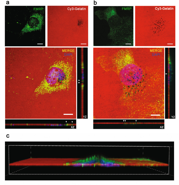
Fig. 4. FMRP is localized inside invadopodia of HuH28 and HuCCT cells.
a A total of 40% confluent HuH28 cells were sown in Cy3-gelatin coated chambers slides and incubated for 19 h for a GDA. Then an IF was performed, and the images were captured using a confocal microscope. Cy3-gelatin is red, FMRP staining green and DAPI staining blue. Representative images of a 60× magnification field (field 15 of 27 total fields). Scale bars: 10 μm. To the right and under the MERGE image, orthogonal views along Y (YZ) and X (XZ) axes are shown, respectively. White arrowheads indicate FMRP staining inside black areas of Cy3-gelatin (degradation areas). b 40% confluent HuCCT cells starved for 24 h in 2% FBS medium were sown in Cy3-gelatin coated chambers slides, treated for 16 h with the MMP inhibitor GM6001 and then incubated for 2 h without MMP inhibition for a GDA. IF and image acquisitions were performed as reported for HuH28. Representative images of a 60× magnification field (field 18 of 28 total fields). Scale bars: 10 μm. To the right and under the MERGE image, orthogonal views along Y (YZ) and X (XZ) axes are shown respectively. White arrowheads indicate FMRP staining inside black areas of Cy3-gelatin (degradation areas). c 3D reconstruction of Z stack (along Y axis) reported in panel (b).

