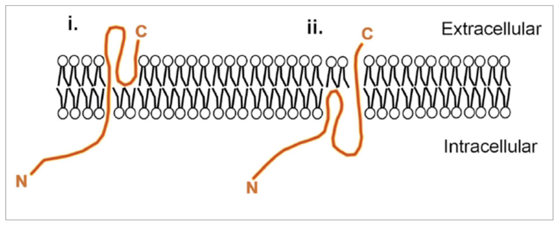
Figure 2. Topology of SynDIG and PRRT proteins.

Schematic of possible models for SynDIG and PRRT topology based on epitope tagging and structural modeling. The loop region between the two predicted membrane segments could be either extracellular (1) as demonstrated for SynDIG1 or intracellular (2) as demonstrated for PRRT2. The topology for SynDIG4/PRRT1 has not been determined. Models are not to scale. Schematic reproduced from Ref. [12].
