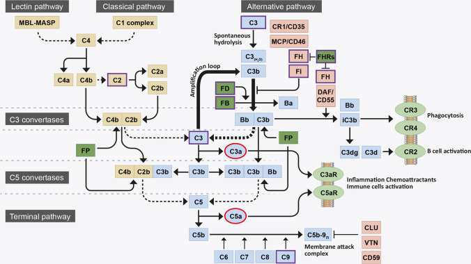
Fig. 1.
Flow diagram of the complement system activation. The complement system is initiated by three activation pathways: the lectin pathway, the classical pathway (both in yellow) and the alternative pathway (indicated in blue). They all converge in the formation of a C3 convertase, responsible for the breakdown (dotted line) of circulating C3 into C3b. During activation via the alternative pathway, FB binds a spontaneously hydrolysed form of C3 [termed C3(H2O)] and is cleaved by FD to form a distinct C3 convertase, C3bBb. This C3 convertase continuously cleaves C3 into C3b, exposing an internal thioester bond within the protein that allows C3b to become covalently attached to local surfaces. Deposited C3b, a potent opsonin, forms the starting block of a surface bound C3 convertase, that cleaves yet more C3 into C3b and contributes to the amplification loop of complement (thick arrows). C3b is also necessary for the formation of a downstream C5 convertase, responsible for the breakdown (dotted line) of C5 into C5b and the subsequent recruitment of C6, C7, C8 and polyC9 into the C5b-9n complex, also known as the membrane attack complement (MAC). The MAC forms a pore on pathogen/cell surfaces and leads to lysis. Complement system activation is tightly controlled by negative (light red) and positive (dark green) regulators: FI and its cofactors FH, CR1 and MCP promote proteolytic cleavage and inactivation of C3b (iC3b) and FH and DAF promote the disassembly of the C3bBb C3 convertase; CLU, VTN and CD59 inhibit the formation of C5b-9n; FP stabilises C3 and C5 convertase; and the FHR proteins compete with FH for C3b binding and inhibit FI-mediated C3b cleavage. C3b breakdown fragments bind membrane-bound receptors (light green): iC3b binds CR3 and CR4 and mediates phagocytosis; C3dg and C3d bind CR2 and mediate B cell activation. Anaphylatoxins C3a and C5a (highlighted in red), generated with the breakdown of C3 and C5, bind membrane-bound receptors (light green) C3aR and C5aR to promote inflammation and immune cells activation. AMD-associated genetic risk variants mostly occur in the genes of the proteins involved in the alternative pathway of complement (highlighted in violet)

