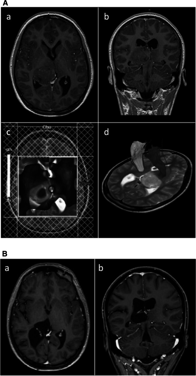
Fig. 2.
(A) Case 2 (a) axial and (b) coronal T1-weighted contrast-enhanced MRI depicting a right posterior thalamic tumor, (c) multivoxel spectroscopy indicating high cellular turnover, (d) DTI depicting the posterior limb of the internal capsule displaced anterolaterally. (B) Case 2 (a) axial and (b) coronal T1-weighted contrast-enhanced MRI at 9 year follow-up after STR via a posterior interhemispheric-subsplenial/parasplenial approach, chemo- and radiotherapy

