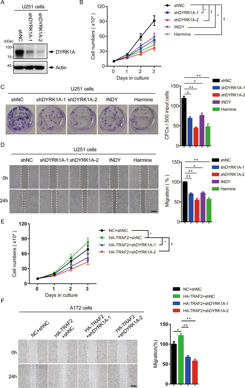
Fig. 7. Depletion of DYRK1A inhibits the growth of glioma cells mediated by TRAF2.
A U251 cells were infected with lentivirus-expressing shRNAs targeting control or DYRK1A. After selection with puromycin for 5 days, cells were lysed and level of DYRK1A analyzed by western blotting. B U251 cells were incubated with 15 μM INDY or 15 μM Harmine, or were infected with shNC or shDYRK1A lentivirus and selected with puromycin. Cell proliferation of the treated U251 cells was measured (n = 3 independent experiments). Data were shown as mean ± SD, *P < 0.05 and **P < 0.01. C Colony-forming capacities of U251 cells treated as in B were measured (CFC, colony-forming cells). Representative images were shown (left) and the quantification of three independent experiments was plotted (right). Data were shown as mean ± SD, *P < 0.05 and **P < 0.01. D The wound-healing assay was used to analyze the migration ability of U251 cells treated as in B. Left panel, representative images were shown. Right panel, the quantification of three independent experiments was plotted. Data were shown as mean ± SD, *P < 0.05 and **P < 0.01, scale bar = 500 μm. E A172 cells were infected with lentivirus-expressing control, HA-TRAF2 and/or DYRK1A shRNAs. Cell proliferation of the A172 cells was measured (n = 3 independent experiments). Data were shown as mean ± SD, *P < 0.05 and **P < 0.01. F The wound-healing assay was used to analyze the migration ability of the treated A172 cells treated as in E. Representative images were shown (left) and the quantification of three independent experiments was plotted (right). Data were shown as mean ± SD, *P < 0.05 and **P < 0.01, scale bar = 500 μm.

