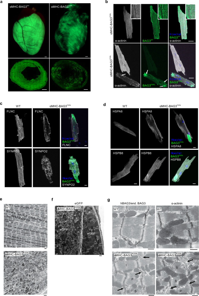
Fig. 1. Characterization of αMHC-BAG3WT and αMHC-BAG3P209L mice.
a Representative hearts and cross-sections from transgenic αMHC-BAG3WT and αMHC-BAG3P209L mice. BAG3WT-eGFP was expressed in the majority of CMs, while BAG3P209L-eGFP showed a patchy expression pattern. Scale bars: 500 µm. b Langendorff-isolated CMs from 10-week-old αMHC-BAG3WT, and αMHC-BAG3P209L mice stained for α-actinin (white) displaying Z-disc localization of BAG3WT (inserts: zoomed regions), but structural disintegration in BAG3P209L expressing CMs (b, arrows). While BAG3WT (green, top panel) formed a grid-like structure (zoomed region, top panel), BAG3P209L (green, bottom panel) was found to form also aggregates of different size (arrows, bottom panel). Scale bar: 20 µm. c, d Langendorff-isolated CMs from 10-week-old WT, and αMHC-BAG3P209L mice were stained for FLNC, SYNPO2 (c), HSPA8, and HSPB8 (d). BAG3P209L (green) formed large aggregates in CMs from αMHC-BAG3P209L mice. Scale bars: 10 µm. e Ultrastructural analysis of hearts from 16-week-old WT and αMHC-BAG3P209L mice; note loss of sarcomeric structures and disarrangement of mitochondria in αMHC-BAG3P209L CMs. Scale bars: 1000 nm. f Immunogold stainings (black dots) of an ultrathin section αMHC-BAG3P209L mice for eGFP. g Immunogold staining for eGFP, hBAG3/endogenous Bag3, and α-actinin with formation of aggregates in CMs from BAG3P209L mice (arrows) accompanied by different stages of sarcomere disintegration. a–g All the experiments were repeated three times from three independent biological replicates with similar results. I, intact sarcomere, P, partly disintegrated sarcomere, S, severely disintegrated sarcomere. WT, wild-type, Scale bars: 1000 nm.

