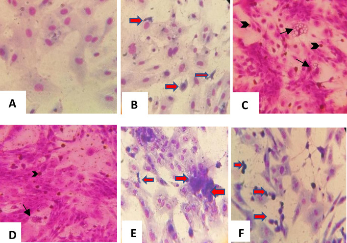
Figure 1.
Giemsa staining of RVFV, SPPV and LSDV viruses for coinfection of LT cell culture (× 400). (A) Mock cells. (B) RVFV cytopathic effect: RVFV necrotic foci (red arrows). (C) SPPV effect (D) LSDV, (E) SPPV/RVFV 0.01/0.01 coinfection of LT cells and (F) LSDV/RVFV coinfection. Capripoxvirus induced cellular damage. Arrows showing capripox virus-induced vacuoles in cells and intracytoplasmic eosinophilic inclusion bodies (Arrowheads).

