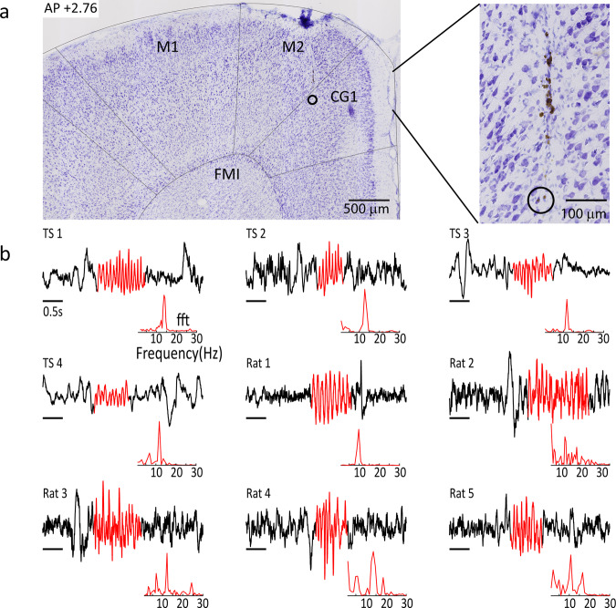
Fig. 6. Validation of recording site and spindle selection.
a Nissl stained section taken ~2.76 mm anterior to bregma from an example rat, showing electrode track and recording site (open circle). b Unfiltered epochs showing examples of spindles detected for each tree shrews and each rat. Red shaded areas indicate the detected spindle, insets below show the fast Fourier transform (fft) of the detected spindle between 5 and 30 Hz. Cg1 anterior cingulate cortex, M1 primary motor cortex, M2 secondary motor cortex, FMI forceps minor corpus callosum.

