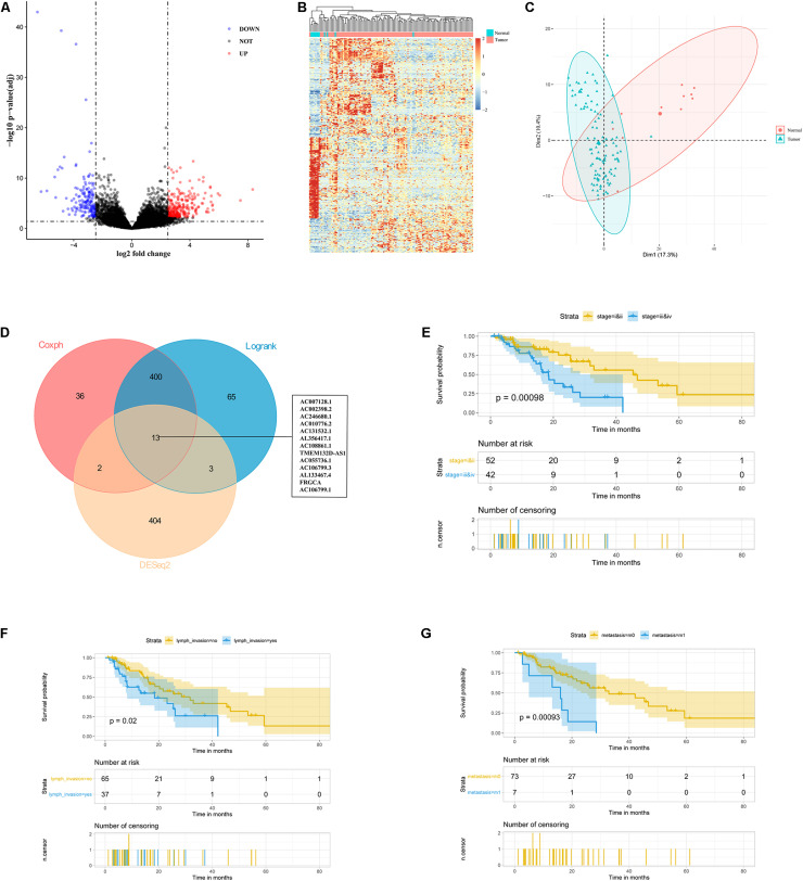
FIGURE 1.
Identification of 13 lncRNAs as candidate prognostic markers for ESCC. (A) Volcano plot exhibited differentially expressed lncRNAs. (B) Heatmap analysis showed that ESCC patients could be distinguished by dysregulated lncRNAs. (C) PCA showed successful segregation between tumor and normal tissues. (D) The intersection of differentially expressed and prognostic lncRNAs by DEseq2, log-rank, and cox analysis. (E–G) Exploratory KM analysis (log-rank test) of the OS probability (with 95% confidence intervals) in ESCC patients [(E) TNM stage; (F) lymph node metastasis; and (G) distant metastasis].

