Figure 4.
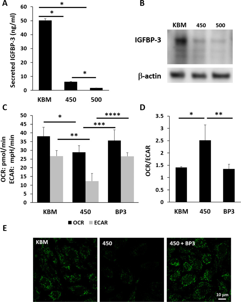
IGFBP-3 blocks the hyperosmolarity-induced decrease in mitochondrial respiration and glycolysis in corneal epithelial cells. The hTCEpi cells were cultured in basal media with or without hyperosmolar conditions for 24 hours. Cells cultured in 450 mOsM were cotreated with 500 ng/mL rhIGFBP-3. (A) IGFBP-3 secretion was down regulated in hyperosmolar culture in basal media (*P < 0.001, one-way ANOVA, SNK multiple comparison test, n = 3). (B) Immunoblotting for intracellular IGFBP-3 in whole cell lysates. Consistent with the decrease in secreted IGFBP-3, intracellular IGFBP-3 was also sequentially decreased with increasing levels of salt. Beta-actin was used as a loading control. (C) Consistent with our prior results, both respiration and glycolysis were significantly decreased after culture in 450 mOsM/kg (**P = 0.021 and ***P = 0.011, one-way ANOVA, SNK multiple comparison test, n = 3). Treatment with rhIGFBP-3 restored metabolism and glycolysis to basal levels (****P = 0.042 and *****P = 0.012, one-way ANOVA, SNK multiple comparison test, n = 3). (D) Hyperosmolar culture shifted cells toward a respiratory phenotype in hyperosmolar culture that was restored following treatment with rhIGFBP-3 (******P = 0.002 and *P < 0.001, one-way ANOVA, SNK multiple comparison test, n = 3). (E) SYBR green staining for mitochondrial DNA showed a decrease in mtDNA after hyperosmolar culture that was blocked by co-treatment with rhIGFBP-3. Scale bar: 10 µm. Number indicates mOsM. Images representative of three repeated experiments.

