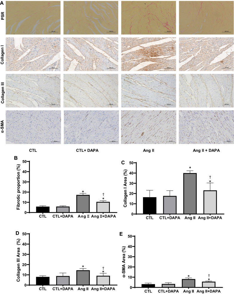
Fig. 3.
DAPA treatment suppresses matrix accumulation and myocardial fibrosis in vitro and in vivo. A The effects of DAPA on the fibrosis in myocardial tissue were observed by PSR staining (top), Scale bars = 100 μm; red color represents collagen fibers deposition. Meanwhile, the expression of type I collagen, type III collagen and α-SMA (bottom) in the myocardium was detected by immunostaining and DAPI staining. Scale bars = 100 μm; B quantification of red color in bar graph. OD values are presented as mean ± SD (n = 6 rats per group). C–E Immunohistochemical analysis for the effect of DAPA on Ang II-induced expression of type I collagen, type III collagen and α-SMA in myocardial tissue. Values are presented as mean ± SD (n = 6 rats per group). *P < 0.05 relative to CTL group. †P < 0.05 relative to Ang II group. F Effects of DAPA treatment on expression of type I collagen, type III collagen, α-SMA in Ang II-infused rats were examined by immunoblotting. The relative ratio of type I collagen, type III collagen, α-SMA over β-actin was determined by densitometric analysis respectively.Values are means ± SD (n = 6). *P < 0.05 relative to CTL group. †P < 0.05 relative to Ang II group. G DAPA inhibits Ang II-induced expression of type I collagen, type III collagen, α-SMA and TGF-β1 in CFs. CFs treated with the indicated concentrations of DAPA for 1 h were exposed to Ang II for 24 h. The relative ratio of type I collagen, type III collagen, α-SMA and TGF-β1 over β-actin was determined by densitometric analysis respectively.Values are means ± SD (n = 3). DMSO dimethyl sulfoxide. Values are means ± SD. *P < 0.05 relative to CTL group. †P < 0.05 relative to Ang II group. §P < 0.05 relative to Ang II plus DAPA 0.5 group


