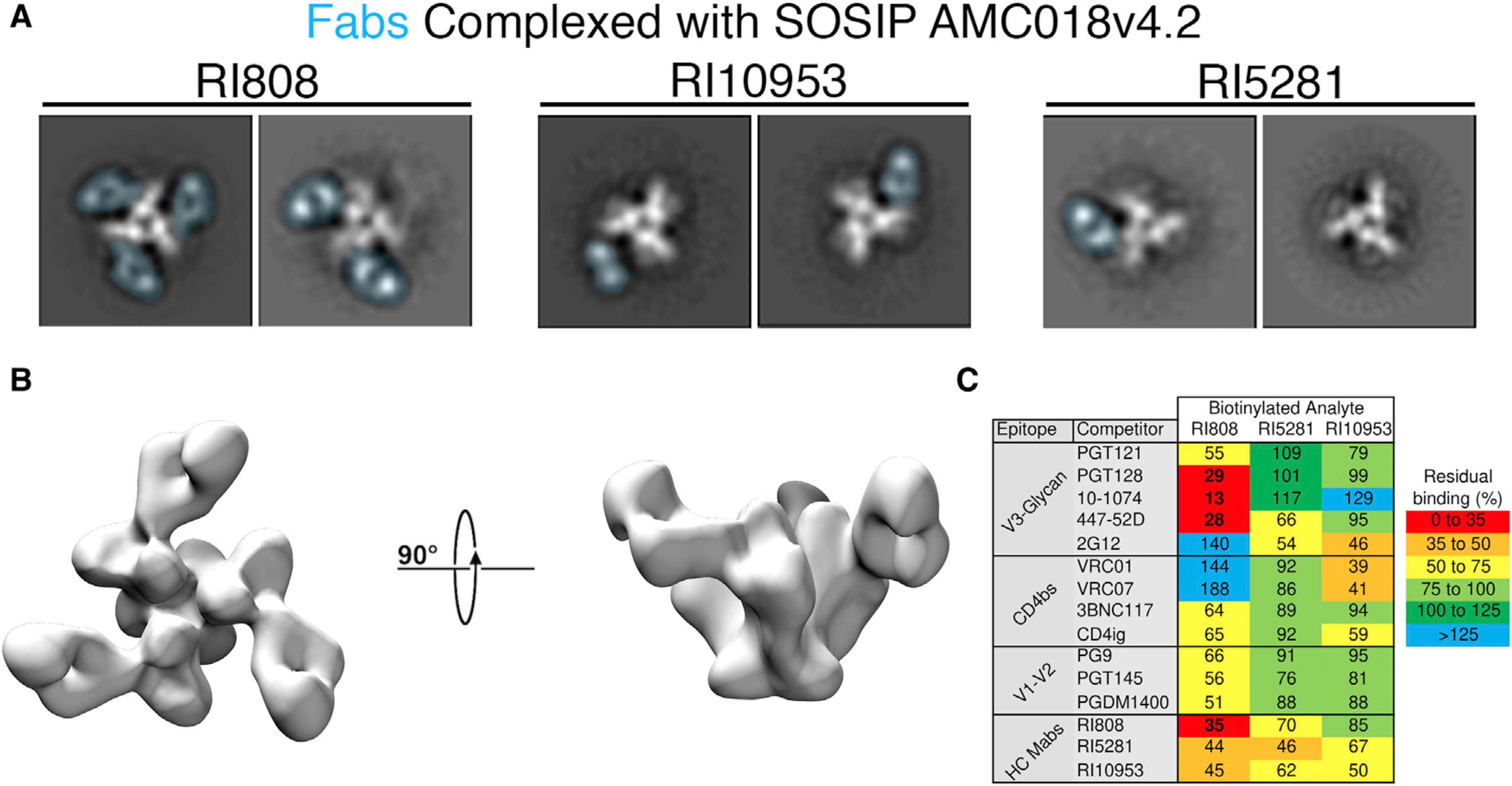
Figure 5. Epitope determination of top HC antibodies.

(A) Negative-stain single-particle analysis of top HC antibodies complexed with SOSIP trimers. 2D Class averages of AMC018 SOSIPv4.2 complexed with the indicated Fab. The density associated with the Fab is overlaid in blue. The structural heterogeneity and low occupancy observed in RI5281 and RI10953 were observed in all 2D class averages.
(B) A 3D negative-stain reconstruction of RI808 complexed with AMC018 SOSIPv4.2.
(C) Competition ELISAs were performed using JR-CSF gp120. The unlabeled competitor is allowed to bind (at 10 μg/ml), followed by the biotinylated antibody being analyzed (at 1 μg/ml). Values indicate %blocked/non-blocked, and numbers indicate the average of triplicate measurements from a representative experiment.
