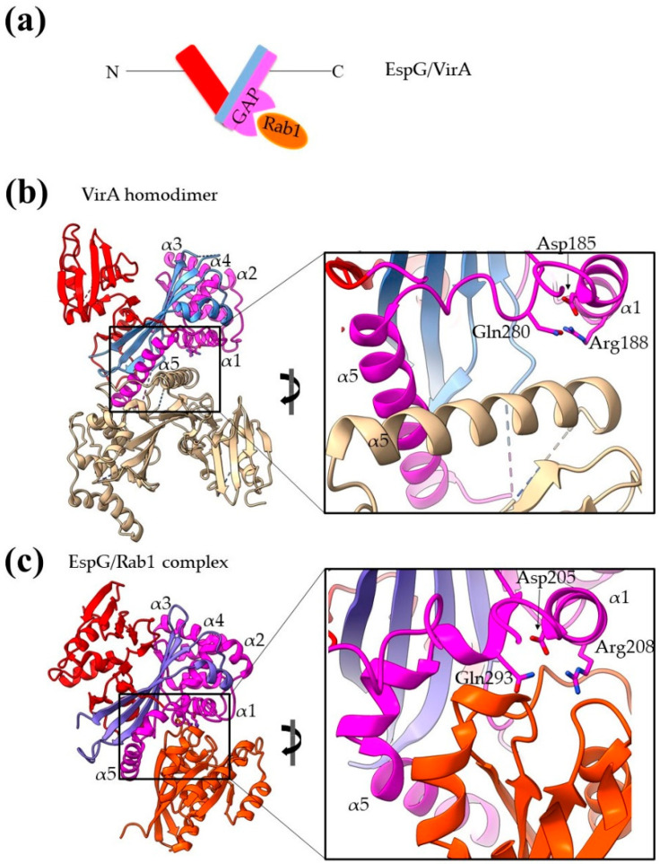
Figure 8.
Bacterial structures with GAP activities. (a) Schematic representation of the domain architecture of EspG/VirA family members. (b) The homodimer of VirA. One monomer is colored white and the other with three different colors highlighting the individual domains. The close-up emphasizes the dimer interface. (c) In the EspG/Rab1 complex, the EspG color code is similar to the one used for VirA, and Rab 1 is colored orange. The close-up emphasizes the complex interaction interface and shows that Rab1 binds on a site equivalent to those used for dimerization. MLD stands for membrane localization domain, SS for secretion sequence, CB for chaperone binding.

