
Figure 1.
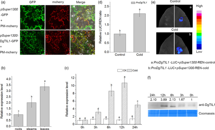
DgTIL1 is responsive to low temperature. (a) Subcellular localization of pSuper1300‐DgTIL1‐GFP in tobacco leaves. pSuper1300‐GFP and CD3‐1007 (an mCherry‐labelled plasma membrane marker) were used as negative controls. Scale bars, 20 μm. (b) Expression of DgTIL1 in the roots, stems, and leaves of WT chrysanthemum at normal temperature using qRT‐PCR (P < 0.05) (Data represent means and standard errors of 3 replicates, 20 plants per replicate). (c) Relative expression levels of the DgTIL1 gene in the WT with low temperature treatment (4°C). CK represents the control under control conditions (25°C day/22°C night). (d and e) DLA assay and LCI assay of the native promoter of ProDgTIL1 after transient expression in tobacco, and the control (25°C day/22°C night) and cold (4°C for 12 h) treatment results were used for comparison. (f) Immunoblot analysis of DgTIL1. Proteins from WT chrysanthemum leaves under low temperature (4°C) treatment were probed with anti‐DgTIL1 (1:1000, from PTM BIO, Hangzhou, China), and Coomassie blue staining was used to demonstrate consistent protein loading.