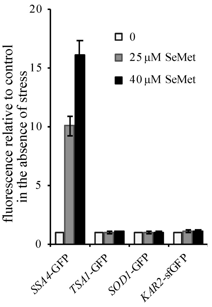
Figure 3.
Cellular stress response to selenomethionine (SeMet) exposure. Exponentially growing BY4741 cells with chromosomally integrated GFP-tagged constructs were incubated in SC + 100 µM methionine for 2 h at 30 °C in the presence of 0 ( ), 25 (
 ) or 40 µM (
 ) SeMet. The fluorescence in whole cell extracts was recorded at 508 nm and normalized to the optical density of the extracts at 280 nm. The fluorescence intensity in the absence of toxic was set as 1. The results are the mean ± S.D. of at least 3 experiments.

