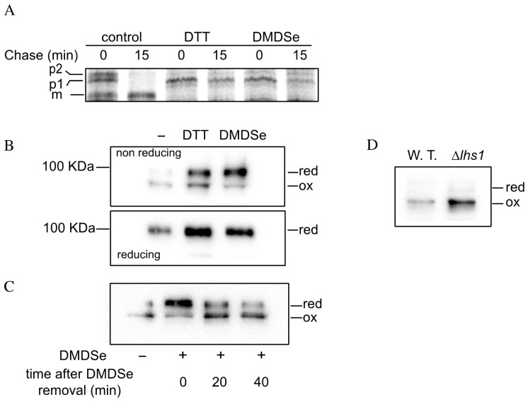
Figure 5.
Disulfide formation in the ER is impaired by exposure to DMDSe. (A) Exponentially growing BY4741 incubated in SC + 100 µM methionine were exposed to nothing (control), 6 mM DTT or 100 µM DMDSe at 30 °C for 20 min before labeling for 10 min with the 35S-Protein mix in a medium without methionine. Cells were harvested after 0 or 15 min of chase with cold methionine and cysteine. Total proteins were extracted and Cpy1p was immunoprecipitated using anti-carboxypeptidase Y antibodies. Immunoprecipitated samples were analyzed by SDS-PAGE (8%), and detected using a Typhoon phosphorimager. The position of the ER (p1) or Golgi (p2) propeptides and the matured protein (m) is indicated. (B) BY4741 cells carrying pAF84 (2 μ ERO1-myc URA3) were grown in SD + 100 µM methionine. Cells were incubated in the absence or presence of 2 mM DTT or 100 µM DMDSe at 30 °C for 30 min. Free thiols were modified with iodoacetamide. Samples, containing equal amounts of proteins, were resolved by nonreducing SDS-PAGE (upper panel) or after the addition of 20 mM DTT in the sample buffer (lower panel). Ero1p-myc was detected by Western blotting with anti-myc antibodies. (C) BY4741 cells carrying pAF84 were incubated in the absence (−) or presence (+) of 50 µM DMDSe for 30 min, after which the incubation was continued in fresh medium without DMDSe. The oxidation state of Ero1p was assessed as in (B) in cell extract harvested either immediately, 20 or 40 min after DMDSe removal. (D) The oxidation state of Ero1p was assessed as in (B), in BY4741 cells (W.T.) or in a ∆lhs1 isogenic strain carrying pAF84.

