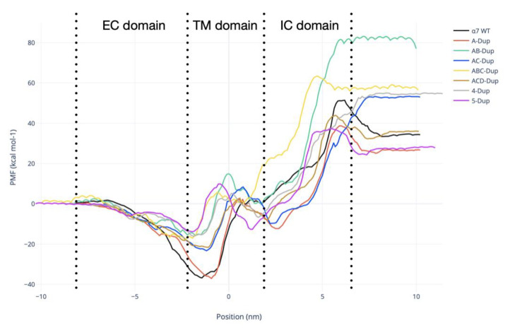
Figure 7.
Potential of mean force (PMF) calculated for the position of the Ca2+ moving through the pentamer axis. The black line shows the PMF obtained for the canonical α7 (WT) receptor, A-Dupα7-red, AB-Dupα7-green, AC-Dupα7-blue, ABC-Dupα7-yellow, ACD-Dupα7-brown, 4-Dupα7-grey, and 5-Dupα7-purple. The schematic arrangements of all models are shown in Figure 8B.

