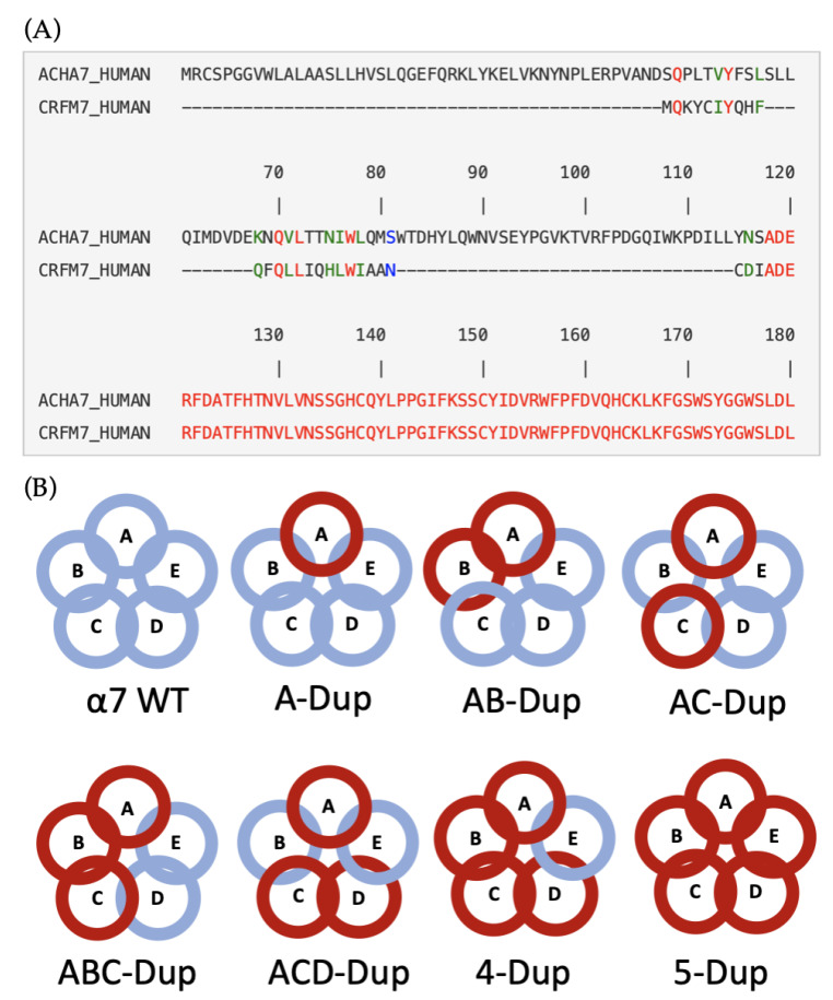
Figure 8.
(A) Sequence alignment of α7/dupα7 extracellular (EC) domains (residues 1–180), performed by ClustalW, green are the residues with high similarity and in red the conserved residues. (B) Schematic representation of all eight different model arrangements dupα7-α7 pentamer, considered in this study: the canonical (WT) α7 subunits are coloured blue; dupα7 subunits are coloured red.

