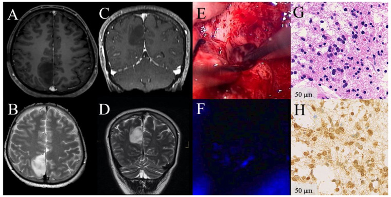
Figure 1.
Illustrative clinical course of a patient with absence of visible 5-aminolevulinic acid (5-ALA) florescence during surgery of a low-grade glioma. Preoperative contrast-enhanced (CE) magnetic resonance images demonstrate a radiologically suspected LGG in the parietal lobe with patchy/faint CE on T1-weighted axial (A) and coronal images (C) and hyperintensity on T2-weighted sequences (B,D). Under white-light microscopy whitish glioma tissue is shown (E). Under violet-blue excitation light, no 5-ALA fluorescence is visible during the entire procedure (F). The corresponding histopathological sample from this non-fluorescing area reveals diffusely infiltrating astrocytoma WHO grade II tissue according to the WHO 2016 criteria in the H&E stain (G). Immunhistochemical staining for IDH mutation is highly positive (H). In the postoperative follow-up, this patient is still alive, more than 11.3 years after initial surgery.

