
Fig. 8.
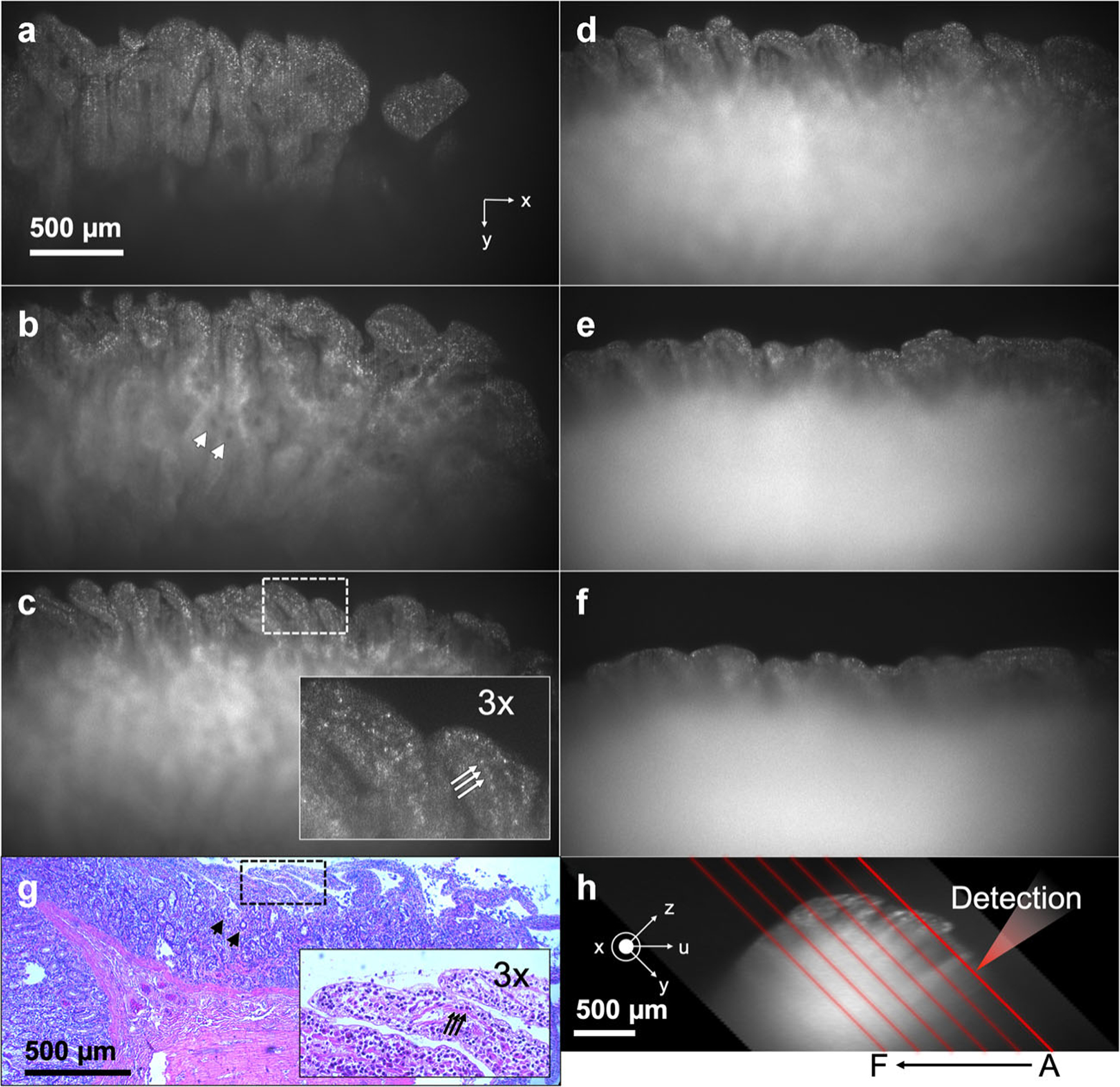
sLSM and histologic images of the excised swine duodenum tissue. (a–f) sLSM images at six different locations as depicted in (h); (g) H&E-stained slide image; and (h) cross-sectional image of the tissue. Arrow, cell nucleus; arrowhead, gland. H&E, hematoxylin and eosin; sLSM, scattering-based light sheet microscope.