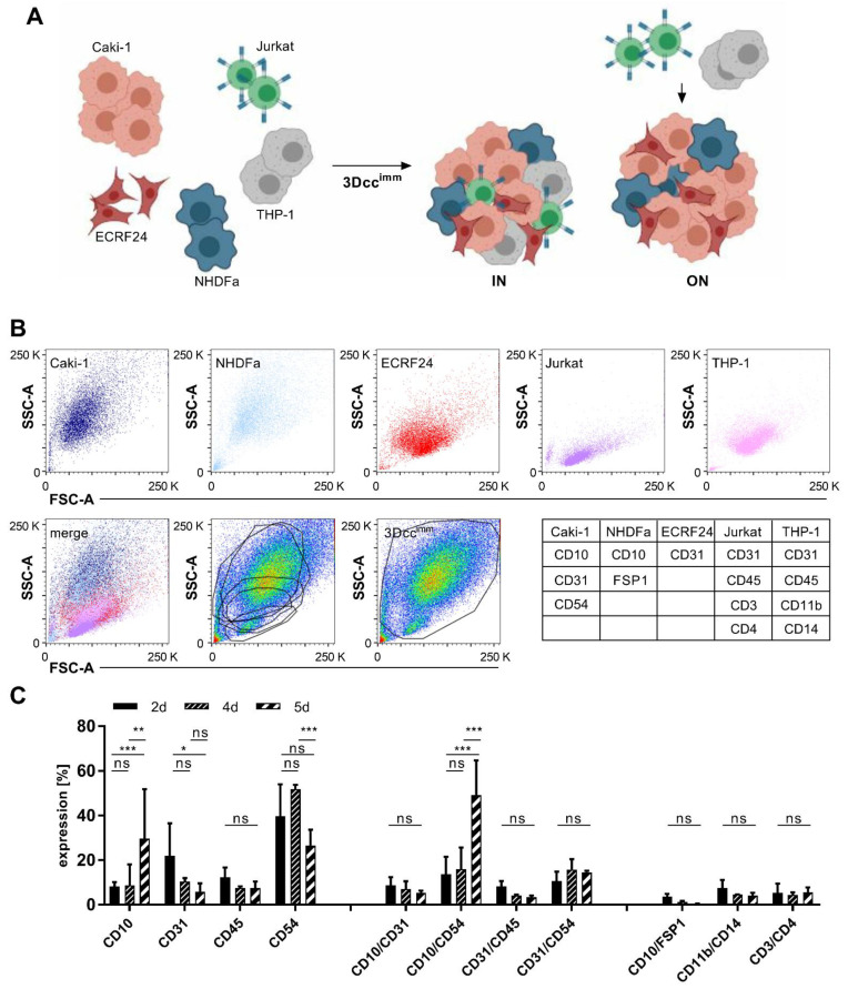
Figure 1.
Formation and maintenance of Caki-1 and Caki-1-SR 3D co-cultures containing immune cells. (A). Scheme of the formation of 3Dccimm spheroids containing ccRCC (Caki-1), endothelial cells (ECRF24), fibroblasts (NHDFα), and immortalized immune cells. To obtain 3Dccimm spheroids, T cells (Jurkat) and monocytes (THP-1) were added at clinically relevant quantities directly during the spheroid formation (IN) or on top of a 24-h pre-formed spheroid (ON). (B). Flow cytometry analysis of the size and granularity (SSC, FSC) of the single cells after 5 days of culturing. Below, an overlay (merge) of the dot blots of the single cells demonstrating the composition of a 3Dccimm spheroid based on the presence of the distinct cell types. Overlay of the single-cell gates onto a pseudocolor blot from dissociated 3Dccimm cultures showing that the size of the single cells changes in the context of the 3Dccimm spheroid and does not allow a precise analysis. Following the global gating strategy (right graph in the bottom panel) single cells in the 3Dccimm were characterized through the expression of distinct cell surface proteins (Table 1). (C). Expression of cell surface proteins in time (2–5d) shown through the FACS analysis. Single and double protein expression has been analyzed in comparison to cell surface proteins exclusively expressed on immune cells. Error bars represent ± SD. Statistical significance was calculated with n = 3 independent experiments by using one-way ANOVA test with unequal variances; * p < 0.05, ** p < 0.01, *** p < 0.001.

