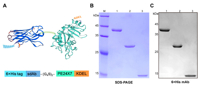
Figure 1.
Characterization of the immunotoxin JVM-PE24X7. (A) An illustrated structure of JVM-PE24X7 constructed by fusing the sdAb (dark blue) with the toxin PE24X7 (mint green). (B) SDS-PAGE and (C) Western blot analysis of purified JVM-PE24X7 (lane 1), PE24X7 (lane 2) and JVM (lane 3). The proteins were loaded onto a 12.5% polyacrylamide gel with protein weight standards and detected by Coomassie brilliant blue staining or Western blot analysis using a mouse anti-His monoclonal antibody.

