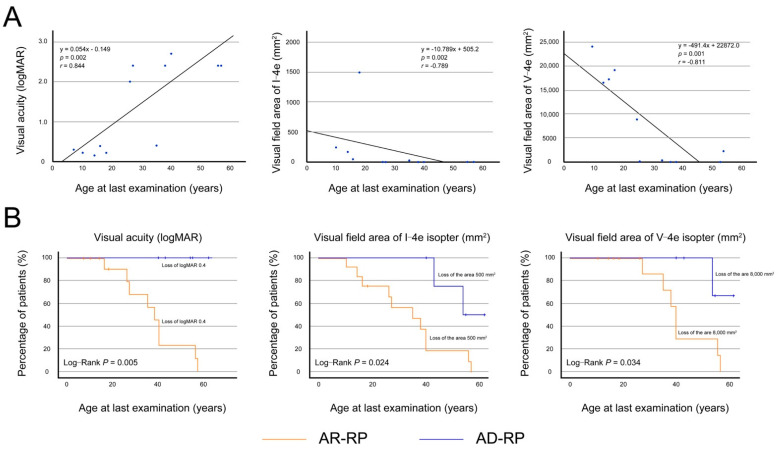
Figure 4.
Visual acuity and visual field areas in patients with autosomal recessive retinitis pigmentosa. (A) In the left eyes (n = 13) of the 13 patients with autosomal recessive retinitis pigmentosa, the graphs show scatter plots of the logarithm of the minimum angle of resolution (logMAR) best-corrected visual acuity (BCVA) and visual field areas of I-4e and V-4e isopters as a function of the age at last examination. There was significant correlation between the BCVA and age (r = 0.844, p = 0.002, Spearman’s rank-order correlation), and between the visual field areas of I-4e and V-4e and age (r = −0.789, p = 0.002; r = −0.811, p = 0.001, respectively). Each graph indicates age-dependent deterioration, reaching to severe impairment around the 50s. (B) The graph shows the Kaplan–Meier survival curves, with log-rank tests, for visual acuity and visual field areas of I-4e and V-4e isopters in patients with autosomal recessive (AR)-retinitis pigmentosa (RP) and autosomal dominant (AD)-RP. The following cutoff points were used: BCVA ≤ 0.4 logMAR units (0.4 decimal units), I-4e isopter area ≤ 500 mm2 (10°), and V-4e isopter area ≤ 8000 mm2 (40°). Patients with AR-RP show significantly faster progression in the loss of visual acuity (p = 0.020) and visual field areas of I-4e (p = 0.011) and V-4e isopters (p = 0.024) in comparison to patients with AD-RP. The survival curves indicate that visual acuity and visual field areas are relatively preserved in most patients with AD-RP until their 40s but are severely impaired in most patients with AR-RP.

