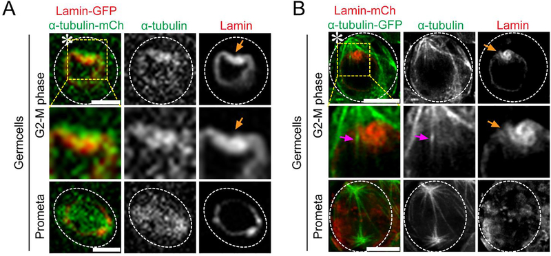
Figure 3: Super-resolution time-lapse imaging of microtubule poking-in activity and asymmetric nuclear envelope breakdown in Drosophila tissue (testes) co-expressing Lamin-mCherry and α-Tubulin-GFP.
(A) Conventional confocal microscopy live cell image showing microtubule dynamics from G2-M phase to mitosis. Image showing higher α-Tubulin-GFP intensity at the stem cell side and its interaction with the nuclear membrane. (B) Airyscan microscope live cell image showing microtubule poking-in and asymmetric NEBD at the stem cell side. Scale bar: 5μm; asterisk: hub (niche); orange arrow: site of microtubule poking in nuclear membrane; magenta arrow: microtubules that poke in at stem cell side.

